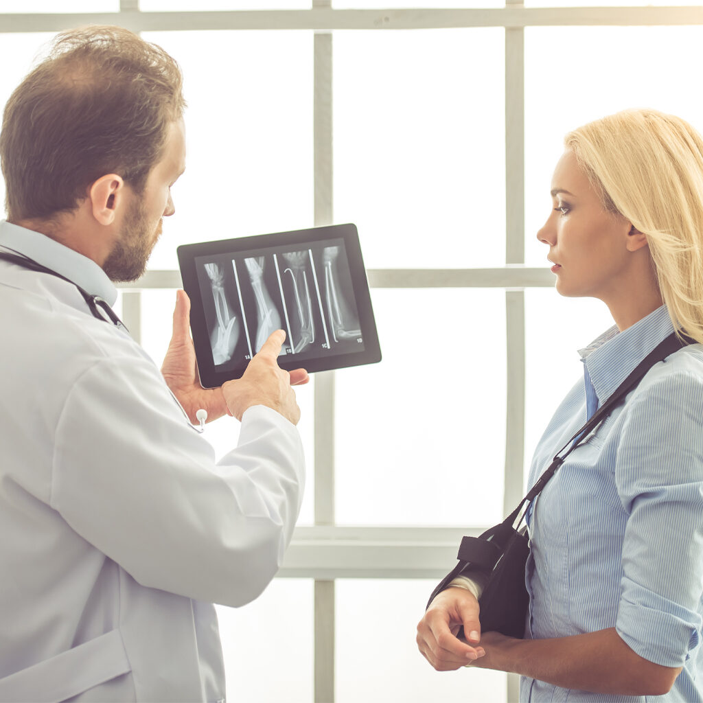
Body/Musculoskeletal Imaging
This subspecialty section consists of radiologists who have expertise in imaging of the chest, abdomen and musculoskeletal system. They specialize in the diagnosis and radiological treatment of various disorders of the kidneys, liver, pancreas, gastrointestinal tract, lung and blood vessels, as well as in orthopedic/bone and joint diseases. New generation CT and MR scanners offer unprecedented three-dimensional detail of anatomy and disease within these body systems, offering non-invasive alternatives to traditional procedures.
Diagnostic Invasive and Therapeutic Procedures
CT, fluoroscopy and ultrasound are the primary imaging modalities used to guide the radiologist in obtaining a biopsy from various organs. The radiologist may choose to perform either core biopsies or fine needle aspirations depending upon the size and location of the lesion to be biopsied. The lungs, liver, pancreas, thyroid, and lymph nodes are among the most commonly biopsied sites. These minimally invasive procedures provide an important alternative to traditional surgical biopsy. Thoracic, abdominal and extremity fluid collections can be sampled, or percutaneous drainage catheters can be inserted, using CT or ultrasound guidance.
These specialists also perform MR, CT and X-ray arthrography of joints, particularly shoulders and hips. Performed with a contrast injection to enhance the visualization of structures within the joints, this procedure aids in the evaluation of joint abnormalities, such as cartilage tears and other injuries.
Non-Invasive Vascular Imaging
Newer MRI and CT technologies allow the radiologist to generate three-dimensional angiographic images of blood vessels within the chest and abdomen. Such non-invasive techniques can be used to evaluate patients with peripheral vascular disease, renal hypertension, mesenteric ischemia, pulmonary embolus and coronary artery disease.
 Skip to content