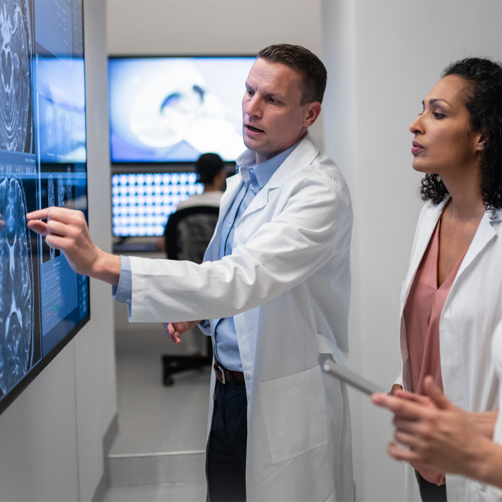
Neuroradiology
Dr. Elliot J. Lerner and Dr. Howard Seigerman are board certified radiologists with additional subspecialty fellowship training in neuroradiology. Both doctors are senior members in the American Society of Neuroradiology, have pursued an accepted course of graduate study and clinical work in neurological disease as outlined by the American Board of Radiology (ABR) and have passed the ABR examination certifying them with added qualifications in Neuroradiology (CAQ).
Dr. Kyung-Hwa Rhee joins the ranks of our neuroradiology team contributing her expertise acquired through extensive fellowship training in spinal imaging.
The demand for expertise solely in the practice of neuroradiology within Bergen County has enabled our doctors to maintain a high standard of excellence in the diagnosis of neurological diseases. State of the art imaging is maintained in a technologically driven medical specialty.
The latest MRI and CT scanners as well as imaging techniques are utilized to diagnose and evaluate neurological diseases, some of which include the evaluation of stroke, tumors, multiple sclerosis, inflammatory/infection conditions, congenital disorders and degenerative spinal
disease.
MRI
Some of the latest innovations utilized include:
- Diffusion Imaging – for early diagnosis of stroke, brain tumors and inflammatory diseases
- Contrast Enhanced MR Angiography – to increase the accuracy for the detection of vascular stenosis and the detection of aneurysms.
CT Imaging
The newest generation of CT Scanners allow us to obtain very high resolution 3-dimensional images of the skull, facial bones and spine. These images play an integral role in clinical decision making as well as pre-and post-operative management.
CT Angiography
Using state of the art CT scanners, neuroradiologists can evaluate for berry aneurysms, vascular stenosis and perform acute stroke evaluation. The latter is particularly valuable for patients unable to undergo MRI evaluation.
Sinonasal Disease
With the ability to use fast scanning, thin section high-resolution images, which are capable of being reformatted in multiple plains, expedites the diagnosis and preoperative evaluation of sinonasal disease. All images will be accessible through a modern digital archiving system. This expedites all forms of communication with the referring clinicians, and reflects our commitment to render the best possible care for our patients.

