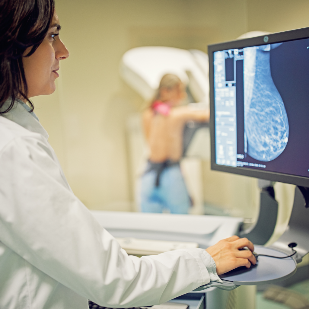
Mammography

Mammography is an X-ray-based imaging test of the breast. It is used by most women preventively to detect breast tumors when they are too small to be detected by physical examination. These small tumors can be the first finding of early-stage breast cancer. Annual mammograms are recommended for all women over 40, and possibly younger if you are at higher risk for breast cancer.

Mammograms are also used to provide better diagnostic detail of a suspicious area within the breast, or to monitor breast cancer or the effects of treatment.

3D mammography (tomosynthesis)

Radiology Associates of Ridgewood has invested in the latest and most advance form of mammography, known as 3D mammography or tomosynthesis. This technology is capable of finding smaller cancers earlier. In addition to improved detection, 3D mammograms have fewer false-positives and fewer call-backs.

The RAR Difference

In addition to being an ACR Designated Comprehensive Breast Imaging Center, RAR offers some of Northern New Jersey’s most experienced breast imaging experts. With state-of-the-art mammography equipment, we believe you shouldn’t have to compromise between accuracy and comfort. That’s why we use the SmartCurve™ paddle system—clinically proven to deliver a more comfortable mammogram compared with standard compression. In fact, it has been shown to improve comfort in 93% of patients who reported moderate to severe discomfort on a previous mammogram.

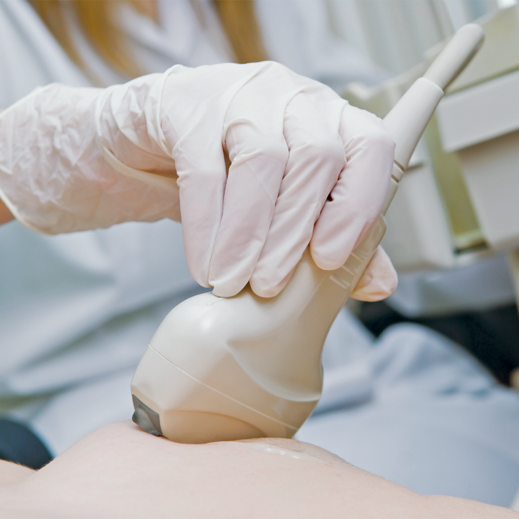
What if I have dense breast tissue? Is an annual mammogram enough?

Dense breast tissue is a risk factor for breast cancer. In addition, breast cancer is more difficult to spot within the fibroglandular tissue of a dense breast. Women with dense breasts may benefit from a supplemental screening option in addition to mammography.

For more information about breast density, talk with your doctor or visit:

Breast MRI

Breast MRI is a non-invasive diagnostic procedure that uses a powerful magnet and radio waves (instead of x-rays) to produce detailed images of the breast. MRI may detect problems within the breast that cannot be seen with a mammogram or ultrasound. 

Breast MRI may be performed with or without and IV injection of contrast (dye) depending upon the indication for the test. MRI does not replace mammography but may be used if additional information is considered necessary by your doctor.

When a breast MRI is needed:

MRI of the breast is not a replacement for mammography or ultrasound imaging but rather a supplemental tool that has many important uses, including:

  • Screening women at high risk, which means a 20 percent or greater lifetime risk of developing breast cancer
  • Determining the extent of breast cancer and to screen the opposite breast after a new diagnosis so the best treatment can be determined
  • Evaluating breast tissue changes during and after treatment for breast cancer
  • Evaluating lumpectomy sites in the years following breast cancer treatment
  • Evaluating breast implants to determine whether silicone implants have ruptured

Before Your Breast MRI

Guidelines about eating and drinking before an MRI exam vary with the specific exam. For some types of exams, you will be asked to fast for 8-12 hours. Unless you are told otherwise, you may follow your regular daily routine and take medications as usual.

Please inform us if you have any serious health problems or if you have recently had surgery. Some conditions, such as severe kidney disease may prevent you from being given contrast material for an MRI. If there is a history of kidney disease, it may be necessary to perform a blood test to determine whether your kidneys are functioning adequately.

You may be asked to wear a gown during the exam or you may be allowed to wear your own clothing if it is loose-fitting and has no metal fasteners.  Jewelry and other accessories should be left at home if possible or removed prior to the MRI scan. Because they can interfere with the magnetic field of the MRI unit, metal and electronic objects are not allowed in the exam room.

If your prior mammograms were not performed at Radiology Associates of Ridgewood, please make them available to us at the time of your exam, if possible.

Breast Ultrasound

A breast ultrasound uses high frequency sound waves to generate images of breast tissues. The sound waves pass through the breast and bounce back, or echo, to form a picture of the internal structures. It is non-invasive and involves no radiation or X-rays. Breast ultrasound is also used to study areas of the breast which can be hard to see with a mammogram, such as the area closest to the chest wall.

When a breast ultrasound is needed:

  • To check a lump felt by a doctor during a clinical exam or by a patient on a self-exam after a mammogram is performed.
  • To further evaluate abnormalities seen on a screening or diagnostic mammogram.
  • To determine whether a breast mass is solid or a fluid-filled cyst.  Solid masses may require follow-up imaging or biopsy.
  • To determine the cause of spontaneous nipple discharge.
  • To evaluate the breast tissue in symptomatic women under age 30.
  • As a supplement to screening mammography in select patients with dense breast tissue.
  • For women who are pregnant and should not be exposed to x-rays.

In most cases, no fasting or special preparation is required before a breast ultrasound. Please dress in clothes that permit access to the area to be tested or that are easily removed.

Dr. Scott DeGregorio and the Importance of Mammograms

You can learn more about the importance of breast cancer screening through an interview with our very own Dr. Scott DeGregorio and CBS News anchor Cindy Hsu.

Contact us today to schedule your appointment!

Skip to content